Sarcomeres are the smallest contractile units of heart and skeletal muscles and are essential for generation and propagation of mechanical force in these striated muscles. During the last decades it has become increasingly clear that components of sarcomeres also play a fundamental role in signal transduction in physiological and pathophysiological conditions. Mutations or misexpression of both sarcomeric contractile and non-contractile proteins have been associated with a variety of cardiac diseases. Moreover, re-expression of foetal sarcomeric proteins or isoforms during cardiac disease can be observed, emphasising the importance of understanding signalling in sarcomeres in both development and disease. The prospective of pharmacological intervention at the level of the sarcomere is now emerging and may lead to novel therapeutic strategies for the treatment of cardiac and skeletal muscle diseases. These aspects will be discussed in this brief review and recent findings, which led to novel insights into the role of the sarcomeric cytoskeleton in muscle development and disease, will be highlighted.
Cardiac and skeletal muscle cells share the presence of myofibrils, highly organised cytoskeletal structures, which are responsible for contractile activity. These myofibrils are composed of repeating contractile units, called sarcomeres. The uniformly arranged thin and thick myofilaments of the sarcomeres give muscle cells their characteristic, microscopically visible, striated pattern. The structure and molecular organisation of sarcomeres are highly conserved between species and show many similarities between cardiac and skeletal muscle cells, despite their distinct functional differences. Henceforth, we will discuss cardiac sarcomeric organisation and function, unless otherwise stated.
Myofibrillogenesis
Studies in developing embryos have made a significant contribution to our current knowledge of myofibril assembly and the importance of different components of the sarcomere, providing us with a molecular basis for a better comprehension of cardiac diseases. The highly orchestrated morphological and molecular events during early cardiac development have been the subject of research in different animal models, such as chick and mouse embryos [1]. After gastrulation during embryonic development, cardiogenic cells from anterior lateral plate mesoderm can be distinguished in the so-called cardiac crescent (first heart field). Later, these cardiogenic cells migrate towards the ventral midline to form the primitive linear heart tube, giving rise to the definitive left ventricle and inflow tract. Cardiogenic cells located medially to the cardiac crescent (second heart field) are recruited during heart tube elongation and looping stages, giving rise to the right ventricle and outflow tract myocardium. Before fusion of cardiogenic cells to the linear heart tube is completed, contractile sarcomeric proteins such as α-actinin are expressed. Alpha-actinin is a major component of the sarcomere and localised at the Z-disc, a structure that defines the boundaries of each sarcomeric unit and thus shared by neighbouring sarcomeres.
Besides the Z-disc, other components of the sarcomere can be distinguished: the I-band (isotropic) surrounds the Z-disc and is composed of only actin filaments, the A-band (anisotropic) is composed of actin and myosin filaments and the M-band (middle) forms the centre of each sarcomere. Contraction is regulated by the coordinated action of the actin-containing thin filaments and the myosin-containing thick filaments, which slide over each other in an ATP-dependent manner. In the heart the giant protein titin is the structural backbone of the sarcomere, indicated by the spanning of the entire Z-disc by two molecules of titin. Titin determines the stiffness of the sarcomere, retains the A-band in the middle of the sarcomere during contraction and is connected with α-actinin at the Z-disc. Alpha-actinin, in its turn, forms homodimers in an antiparallel manner and cross-links actin.
Mutations in sarcomeric proteins are associated with cardiomyopathies
Cardiomyopathy is a group of disorders of the heart that result in inadequate pumping of blood around the body. Hypertrophic cardiomyopathy (HCM) and dilated cardiomyopathy (DCM) are mainly caused by mutations in sarcomeric proteins. HCM is characterised by left ventricular hypertrophy (resulting from increased cell size or volume), increased systolic function and decreased diastolic function. At the histological level, hypertrophy and disarray of cardiomyocytes and interstitial fibrosis is observed. DCM, on the other hand, is characterised by ventricular dilation and contractile dysfunction of the left and/or right ventricles. Depending on the location of the mutation, HCM or DCM is induced; in HCM regions of sarcomeric proteins are mutated that are directly involved in force transmission, whereas the regions that are mutated in DCM are involved in force transmission from the sarcomere to the cytoskeleton. The majority of mutations in sarcomeric proteins are found in myosin heavy chain β and myosin binding protein C [2, 3].
Besides mutations in proteins that affect force generation and transmission, mutations in proteins at the Z-disc (and also the M-band) that disrupt stress sensing and downstream signalling have also been implicated in cardiomyopathies and severe hypertrophic responses. For instance, calcineurin, muscle-specific LIM protein (MLP), T-cap, calsarcin-1, cypher and Enigma homologue protein (ENH) interact with α-actinin at the Z-disc and have been associated with cardiomyopathies [4]. The putative stress sensor MLP has been shown to be an essential component of the T-cap and titin complex, stabilising the interaction between these proteins, which can then sense an increase in stretch, followed by activation of downstream signals for the induction of hypertrophy [5]. The calcium-dependent phosphatase calcineurin is a key regulator of hypertrophy. Activated calcineurin dephosphorylates nuclear factor of activated T-cells (NFAT), which subsequently translocate to the nucleus to activate genes that are associated with stress, such as atrial natriuretic factor (ANF) and brain natriuretic peptide (BNP), and genes that encode foetal isoforms of contractile proteins, such as β-myosin heavy chain (β-MHC). Calcineurin transgenic (Tg) mice, expressing a constitutively active form of the catalytic subunit of calcineurin in the heart, show massive hypertrophy, disorganised cardiomyocytes, fibrosis and enlarged nuclei.
Calsarcin, a novel target for calcineurin, binds to both calcineurin and α-actinin. Overexpression of calsarcin-1, both in vitro and in vivo, has protective effects on hypertrophy by interfering with calcineurin-NFAT signalling, while a lack of calsarcin-1 sensitises the calcineurin pathway and shows accelerated formation of cardiomyopathy in response to pathological stimuli. Cypher (or Oracle/ZASP) is expressed in heart and skeletal muscle during mouse and zebrafish development and interacts with α-actinin and calsarcin. Deletion of cypher in mice and zebrafish resulted in disruption of the Z-disc structure leading to DCM. However, in mice only deletion of the long isoform of cypher led to development of late-onset DCM, whereas deletion of the short isoform had no effect. More interestingly, mutations in cypher that cause DCM have been identified in humans. These results indicate cypher as an essential component of the α-actinin-calcineurin-calsarcin complex in order to maintain cardiac structure and function. ENH is another PDZ-LIM protein, which interacts with α-actinin, calsarcin and cypher. In ENH−/− mice expression of the short cypher isoform and calsarcin is lost, which leads to disruption of the Z-disc and development of DCM. These data show an essential role for the Z-disc in the development of cardiomyopathies and targeting these components could reveal strategies for possible new therapies for the treatment of these diseases.
Actin signalling and transcriptional regulation in cardiac development and disease
The actin cytoskeleton in striated muscles is organised in sarcomeres for generation of contractile force, as we have previously discussed. Actin is also important in other cellular processes, including cytokinesis, migration, regulation of cell shape and gene expression during development and disease. In recent years, it has become clear that regulators of actin dynamics are crucial for the assembly and maintenance of functional sarcomeres [6]. Polymerisation of monomeric globular actin (G-actin) in filamentous actin (F-actin) is regulated by several actin-binding proteins. Rho GTPases (RhoA, Rac and Cdc42) induce actin polymerisation via the Rho-associated kinase (ROCK)-LIM kinase-cofilin pathway. RhoA is highly expressed in embryonic hearts, down-regulated in adult hearts and re-expressed during cardiac hypertrophy. Heart-specific overexpression of RhoA leads to the development of a DCM phenotype, whereas inhibiting ROCK protects the heart after aorta constriction. In addition, actin signalling has an important role in the regulation of transcriptional activity of various transcription factors, such as serum response factor (SRF), myocardin-related transcription factors (MRTFs) and MEF2.
SRF is a member of the MADS (MCM1, Agamous, Deficiens, SRF) family of transcription factors, which share a conserved motif of 57 amino acids mediating homodimerisation, association with other factors and DNA-binding activity. Homodimeric SRF binds to a palindromic DNA sequence (CC(A/T)6GG), which is called the CArG box. Several SRF-mouse models have shown that SRF is essential for cardiac development, as well as maintaining adult cardiomyocytes. SRF is responsible for maintaining the sarcomeric integrity in cardiomyocytes and disruption of its expression results in DCM in mice. Therefore, it is not surprising to find several sarcomeric genes, such as skeletal and cardiac actin, α-MHC and β-MHC, myosin light chain (MLC)2a and 2v, myomesin, troponin C, and titin, to be targets of SRF [7].
Myocardin, a co-activator of SRF, is essential for smooth muscle differentiation and activates expression of smooth muscle genes in promoters with two adjacent CArG boxes by binding these as homodimers. Cardiac specific deletion (using an α-MHC-Cre mouse line) of myocardin leads to decreased expression of sarcomeric genes (α-cardiac actin, MLC2v, and tropomyosin) and connexin 43 and desmin, which results in disruption of the structural organisation of the sarcomere and intercalated discs, respectively. Furthermore, the hearts of α-MHC-Cre-myocardinf/f showed left atrial enlargement, hypertrophy, interstitial fibrosis and a decreased cardiac function (decreased ejection fraction). Myocardin also has an essential function during pathophysiological events in the heart; it is upregulated in several in vivo and in vitro models of hypertrophy, as well as in human patient hearts with idiopathic DCM. The hypertrophic marker ANF is a direct target of myocardin and overexpression of myocardin in cardiomyocytes in vitro leads to a hypertrophic response, which can be abolished by expression of a dominant negative isoform lacking the transcriptional activation domain.
Myocyte enhancer factor-2 (MEF2) is another MADS-box transcription factor. Four isoforms of MEF2 exist (A–D), of which isoforms A, C and D have distinct functions in the heart. Knock-out studies show that MEF2A is essential for energy metabolism and sarcomeric organisation in adult hearts, MEF2C is involved in cardiac morphogenesis, and MEF2D is a mediator of stress-dependent pathological remodelling of the adult heart. On the other hand, overexpression of these factors induced (dilated) cardiomyopathy in mice.
Transcriptional activity of SRF is regulated by Striated Muscle Activator of Rho Signalling (STARS). STARS binds to actin and activates RhoA, which promotes actin polymerisation and depletion of the G-actin pool. Subsequently, MRTFs translocate to the nucleus (via decreased binding to G-actin binding domain) and activate gene expression by acting as co-factor for SRF. SRF induces actin genes, in this way providing a negative feedback loop. STARS has been shown to be a direct target of MEF2 in different models of hypertrophy [7]. Thus, the formation of actin bundles is not only necessary for cell movement and polarisation, but also leads to the induction of signal-transduction pathways important for cardiac development and hypertrophy.
CHAP, a sarcomeric protein, important for cardiac development and disease
Recently, following whole genome-wide microarray analysis of pluripotent human embryonic stem cell differentiating towards functional cardiomyocytes, we identified a novel gene, which we named Cytoskeletal Heart-enriched Actin-associated Protein (CHAP) (also known Synaptopodin-2-like (SYNPO2L)) and which is highly expressed in heart and skeletal muscle [8]. CHAP displays a significant homology to myopodin and synaptopodin (31 % and 30 %, respectively), which are both actin-bundling proteins that bind to α-actinin. Whereas synaptopodin is expressed in kidney and forebrain, myopodin is predominantly expressed in skeletal muscle and at lower levels in heart and smooth muscle cells. Interestingly, myopodin is localised at the Z-disc of skeletal muscle cells and cardiomyocytes, but can be translocated to the nucleus in a differentiation- and stress-dependent manner [9]. Phosphorylated synaptopodin and myopodin, mediated by inhibition of calcineurin activity, resulted in stabilisation of the actin cytoskeleton in kidney podocytes [10] and translocation from the Z-disc to the nucleus in cardiomyocytes [11], respectively.
Based on the homology of CHAP with both myopodin and synaptopodin, we decided to further explore the role of CHAP in cardiomyocyte differentiation and more specifically in sarcomere organisation and function. We identified two isoforms of human and mouse CHAP: a long isoform CHAPa and a shorter isoform CHAPb. CHAPa and CHAPb isoforms are almost identical, containing a putative nuclear localisation signal (NLS) and actin binding sites. The N-terminal of CHAPa contains an additional PDZ domain. In chick one CHAP isoform has been identified, which shows homology with the human and mouse CHAPa gene [12]. During mouse embryonic development, ChapB is first expressed at the cardiac crescent stage (E7.75), later in the linear heart tube and in the looped heart. From embryonic day 10 onward, expression is also detected in somites (giving rise to skeletal muscle). Expression of ChapB is downregulated in adult hearts, whereas ChapA is expressed in adult heart and skeletal muscle [13]. Furthermore, Chap is expressed in developing muscles, such as jaw, tongue and eye muscle. A similar expression pattern of CHAP in chick embryos was observed [12]. In addition, we showed expression of mouse Chap in skeletal muscle and smooth muscles of the aorta, carotid artery and coronary arteries. These results suggest a function for CHAP in muscle development and adult muscle.
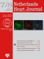
Similar to myopodin, we demonstrated that CHAP was localised at the Z-disc of cardiomyocytes and skeletal muscle cells, supported by co-localisation of α-actinin and the lack of overlap in localisation with the M-band marker myomesin (Fig. 1). Moreover, besides their co-localisation, we also showed that CHAP was able to interact with α-actinin-2. In a next step we studied the role of CHAP during zebrafish development. Morpholino-mediated knock down of chap in zebrafish resulted in defects in heart looping, cardiac oedema and disturbed skeletal muscle development [13]. These data indicated an essential role for CHAP in cardiac and skeletal muscle development.
Fig. 1
CHAP is localised at the Z-disc. Mouse embryonic stem cells were differentiated to cardiomyocytes and immunostained with antibodies for the Z-disc protein CHAP (green, left), M-band marker myomesin (red, middle). Nuclei are stained with TOPRO-3 (blue). In the merge panel (right) it can be appreciated that CHAP and myomesin do not co-localise
×
In order to investigate the role of CHAP in adult mice, we generated transgenic mice by heart-specific overexpression of both isoforms of CHAP. CHAPb transgenic mice displayed a phenotype, which resembles dilated cardiomyopathy and was associated with abnormal atrial conduction (unpublished data). Interestingly, in a recent large-scale meta-analysis of multiple genome-wide association studies of European ancestry six new susceptibility loci for atrial fibrillation were identified, of which one locus was located 5 kb upstream of SYNPO2L (CHAP) and 20 kb upstream of MYOZ1 (calsarcin-2) [14]. These findings corroborate the changed atrial conductivity in CHAP Tg mice.
Clinical perspective
It is evident that well-coordinated activities of the various components of the sarcomere are necessary for proper function of the heart muscle. Changes in expression levels, localisation or function of these components may ultimately lead to heart failure. Heart failure patients with impaired cardiac contractility can be treated with drugs that enhance β-receptor cAMP-dependent protein kinase pathways, leading to increased phosphorylation of many different target proteins (including sarcomeric proteins) and increased cardiac contractility. However, detrimental side effects can be observed, because of the lack of selectivity of these drugs. Nevertheless, recent successful development of selective activators of sarcomeric contractile and their regulatory proteins (i.e. kinases) demonstrate that the sarcomere is a potentially promising drug target [15].
In addition, significant recent advances in (human) pluripotent stem cell biology now enable efficient generation of functional cardiomyocytes in vitro from patients and healthy individuals [16]. Studying abnormal cardiomyocyte function at the cellular level may provide a better understanding of the underlying mechanism(s) responsible for the cardiac disease phenotype in patients. Moreover, implementation of in vitro cardiac disease models for drug discovery may be valuable for screening and identification of effective and selective drugs, leading to improved therapeutic strategies.
In conclusion, the perception of the sarcomere has considerably changed during the last two decades, going from a static contractile unit, responsible for generation and propagation of mechanical force, to a dynamic unit which, in addition to its essential role in contractility, is important for sensing mechanical stress or other changes in upstream signalling and the translation to adequate downstream signalling events. An increased knowledge in sarcomeric biology and function will be fundamental for unravelling signalling pathways that are important for both muscle development and disease. Moreover, recent advances in pharmacological interventions with small molecule drugs have shown functional responses at the level of individual myosin and actin filaments, leading to an increased awareness of the potential of sarcomeric proteins as pharmacological targets. Ultimately, this may lead to new therapeutic strategies for the treatment of a variety of muscle diseases.
Acknowledgments
This study was supported by the Netherlands Heart Foundation grant 2006B209 (W.E.).
Conflict of interests
None declared.
Open Access This article is distributed under the terms of the Creative Commons Attribution License which permits any use, distribution, and reproduction in any medium, provided the original author(s) and the source are credited.
Het Netherlands Heart Journal wordt uitgegeven in samenwerking met de Nederlandse Vereniging voor Cardiologie en de Nederlandse Hartstichting. Het tijdschrift is Engelstalig en wordt gratis beschikbaa ...