The use of biplane fluoroscopy to track bones in the foot is challenging, due to distortion, overlap and image artefact inherent in fluoroscopy systems and high speed photography. The accuracy and precision of these systems have been reported [1‐4] and are presented here for our biplane fluoroscopy system.
Materials and methods
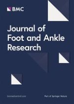
Biplane Fluoroscopy System: The system consists of two Philips BV Pulsera C-arms set in custom frames around a raised floor with a radiolucent imaging area. X-ray images are captured with high speed (1000fps) cameras. Validation Object: 1.6mm tantalum beads were placed in a machined block (wand) then measured to 7 microns with a Coordinate Measuring Machine to determine their centroid location. The wand was translated and rotated via a 1 micron precision stepper-motor for static validation, as well as manually swept through the field of view at ~0.5m/s for dynamic. Static Accuracy and Precision: accuracy was defined as the RMS error between the translation of the stepper-motor and the measured movement of the beads; precision is defined as the standard deviation of the bead locations. For rotation, accuracy was defined as the RMS error between the applied and measured rotation of the wand. Dynamic Accuracy and Precision: accuracy was defined as the RMS error between the known and measured inter-bead distance; precision was the standard deviation of the inter-bead distance. 3D location processing was accomplished using custom software written in MatLab to derive the 3D location of objects from two, time-synchronized, 2D fluoroscopy images of known spatial relationship. This software also compensates for the image distortion (Figure 1).
Figure 1
Distortion plate before (left) and after (right) correction for pin-hole distortion and magnetic distortion.
×
Results
Translation: the overall RMS error was 0.066 mm, with a precision of ± 0.016 mm. Rotation: the RMS error was 0.125°. Dynamic motion: the overall RMS error was 0.126 mm, with a precision of ± 0.122 mm.
Conclusions
The accuracies and precision in the results are comparable to similar such systems in development to investigate other joints of the body [1‐4]. We are currently developing and validating a marker-less technique for tracking the bones of the foot.
Open Access
This article is published under license to BioMed Central Ltd. This is an Open Access article is distributed under the terms of the Creative Commons Attribution License (
https://creativecommons.org/licenses/by/2.0
), which permits unrestricted use, distribution, and reproduction in any medium, provided the original work is properly cited.
Binnen de bundel kunt u gebruik maken van boeken, tijdschriften, e-learnings, web-tv's en uitlegvideo's. BSL Podotherapeut Totaal is overal toegankelijk; via uw PC, tablet of smartphone.