Abstracts
The aim of this research was to evaluate dental age in 102 patients with Down Syndrome, using panoramic radiographs. A software program developed by the Discipline of Radiology, School of Dentistry of São José dos Campos, São Paulo State University (UNESP), was used. A table of mineralization chronology of permanent teeth among Brazilians conceived by Nicodemo, Moraes and Medici Filho was used within the software. Statistical analysis of the results showed that 70.91% of the males and 61.21% of the females presented advanced dental age. Only 32.09% of the males and 38.79% of the females presented delayed dental age. Regarding the differences between the dental and chronological ages, two thirds of the males and females presented dental age with differences of up to 12 months, which means that they can be considered to be within normal standards, whereas only 18.87% of the males and 10.21% of the females presented dental age outside normal standards, with differences of over 24 months. In conclusion, the majority of the patients with Down Syndrome were considered to be within the normal standards of mineralization chronology.
Down syndrome; Age determination by teeth; Tooth calcification; Radiography, panoramic
O objetivo desta pesquisa foi avaliar a idade dentária em 102 indivíduos com síndrome de Down, por meio de radiografias panorâmicas. Foi usado um "software" desenvolvido pela disciplina de Radiologia da Faculdade de Odontologia de São José dos Campos (UNESP). Neste "software", foi utilizada a tabela de cronologia de mineralização dos dentes permanentes entre brasileiros, de Nicodemo, Moraes e Medici Filho. A análise estatística dos resultados mostrou que 70,91% dos indivíduos do sexo masculino e 61,21% do sexo feminino apresentaram idade dentária adiantada. Apenas 32,09% dos indivíduos do sexo masculino e 38,79% dos indivíduos do sexo feminino apresentaram idade dentária atrasada. Com relação às diferenças entre idade dentária e idade cronológica, dois terços dos indivíduos dos sexos masculino e feminino apresentaram idades dentárias com até 12 meses de diferença, o que significa que estão dentro do padrão de normalidade; enquanto 18,87% dos indivíduos do sexo masculino e 10,21% dos indivíduos do sexo feminino apresentaram idades dentárias fora do padrão de normalidade com diferenças acima de 24 meses. Concluiu-se que a maioria dos pacientes com síndrome de Down estava dentro do padrão de normalidade de desenvolvimento dentário.
Síndrome de Down; Determinação da idade pelos dentes; Calcificação de dente; Radiografia panorâmica
ORIGINAL ARTICLES
RADIOLOGY
Dental age in patients with Down syndrome
Idade dentária em pacientes com síndrome de Down
Mari Eli Leonelli de MoraesI; Michelle Silva BastosII; Luis Roque de Araujo dos SantosIII; Julio Cezar de Melo CastilhoIV; Luiz Cesar de MoraesV; Edmundo Medici FilhoV
IPhD, Professor Department of Diagnosis and Surgery, School of Dentistry of São José dos Campos, São Paulo State University
IIScientific Initiation Student Department of Diagnosis and Surgery, School of Dentistry of São José dos Campos, São Paulo State University
IIIPhD Student Department of Diagnosis and Surgery, School of Dentistry of São José dos Campos, São Paulo State University
IVAdjunct Professor Department of Diagnosis and Surgery, School of Dentistry of São José dos Campos, São Paulo State University
VChair Professors Department of Diagnosis and Surgery, School of Dentistry of São José dos Campos, São Paulo State University
Corresponding authorCorresponding author:Luis Roque de Araujo dos Santos Rua Santa Rita de Cássia, 60, Vila Japão Itaquaquecetuba - SP - Brasil CEP: 08599-010 E-mail: luisroquearaujo@hotmail.com
ABSTRACT
The aim of this research was to evaluate dental age in 102 patients with Down Syndrome, using panoramic radiographs. A software program developed by the Discipline of Radiology, School of Dentistry of São José dos Campos, São Paulo State University (UNESP), was used. A table of mineralization chronology of permanent teeth among Brazilians conceived by Nicodemo, Moraes and Medici Filho was used within the software. Statistical analysis of the results showed that 70.91% of the males and 61.21% of the females presented advanced dental age. Only 32.09% of the males and 38.79% of the females presented delayed dental age. Regarding the differences between the dental and chronological ages, two thirds of the males and females presented dental age with differences of up to 12 months, which means that they can be considered to be within normal standards, whereas only 18.87% of the males and 10.21% of the females presented dental age outside normal standards, with differences of over 24 months. In conclusion, the majority of the patients with Down Syndrome were considered to be within the normal standards of mineralization chronology.
Descriptors: Down syndrome; Age determination by teeth; Tooth calcification; Radiography, panoramic.
RESUMO
O objetivo desta pesquisa foi avaliar a idade dentária em 102 indivíduos com síndrome de Down, por meio de radiografias panorâmicas. Foi usado um "software" desenvolvido pela disciplina de Radiologia da Faculdade de Odontologia de São José dos Campos (UNESP). Neste "software", foi utilizada a tabela de cronologia de mineralização dos dentes permanentes entre brasileiros, de Nicodemo, Moraes e Medici Filho. A análise estatística dos resultados mostrou que 70,91% dos indivíduos do sexo masculino e 61,21% do sexo feminino apresentaram idade dentária adiantada. Apenas 32,09% dos indivíduos do sexo masculino e 38,79% dos indivíduos do sexo feminino apresentaram idade dentária atrasada. Com relação às diferenças entre idade dentária e idade cronológica, dois terços dos indivíduos dos sexos masculino e feminino apresentaram idades dentárias com até 12 meses de diferença, o que significa que estão dentro do padrão de normalidade; enquanto 18,87% dos indivíduos do sexo masculino e 10,21% dos indivíduos do sexo feminino apresentaram idades dentárias fora do padrão de normalidade com diferenças acima de 24 meses. Concluiu-se que a maioria dos pacientes com síndrome de Down estava dentro do padrão de normalidade de desenvolvimento dentário.
Descritores: Síndrome de Down; Determinação da idade pelos dentes; Calcificação de dente; Radiografia panorâmica.
Introduction
Patients with Down syndrome have a chromosomal aberration resulting from a trisomy of the 21st. The most likely cause is non-disjunction occurring during gametogenesis (meiosis). It carries this name because the syndrome was first studied by an English physician called John Langdon Down, in 1866.1,4,9,13
The Down syndrome involves a set of signs and symptoms that characterize a delay in the development of the motor and mental functions of its carriers, entailing mental and general alterations.2,8,11,15
The following general alterations are involved:
1. eyes - slanting, almond-shape, plica palpebronasalis (epicanthus), strabismus, myopia;
2. nose - flattening of the nose bridge, small, short nose with broad nasal bridge, pug nose;
3. ears - lop ears with flat or absent helix, auricles with a low implantation;
4. wide and short neck with abundant skin;
5. congenital cardiopathy;
6. wide hands;
7. short fingers;
8. clinodactyly;
9. brachydactyly;
10. muscular hypotony.
Delay occurs in the pre-natal and post-natal development, and weight and stature values are observed to be generally below the normal standards.2,3,6,8,17
Regarding mental aspects, patients with Down Syndrome present alterations in motor coordination and intelligence. According to Coelho, Loevy2 (1982), the average intelligence quotient (IQ) for Down patients is 36.5.
According to Mariano et al.5 (1999), Pilcher10 (1998) and Ribeiro et al.12 (2003), the systemic alterations with special dental significance in Down Syndrome patients are heart disease, pneumonia, allergy, bronchitis, tonsillitis, convulsion and blood dyscrasia.
Upon analyzing the chronological development of permanent teeth of Down children, Toscano17 (1994) found a delay in the maxilla and mandible in males and females.
The following dental alterations are involved:
periodontal disease - it is a universal finding, with an incidence of 90-96%;
caries - reduction of caries when compared to controls;
increased incidence of malocclusion, particularly Class III malocclusions;
dental anomalies, particularly anodontia;
delayed eruption of the permanent teeth;
impacted teeth, particularly among cuspids andbicuspids;
macroglossia, with an incidence of 11-60%.
Santos14 (2004) studied the mineralization chronology in patients with Down Syndrome using panoramic radiographs. Mineralization evaluation of the panoramic radiographs was performed according to the stages proposed by Nicodemo, Moraes and Medici Filho, and a delay was found in the development of the canines and second molars. Sannomiya et al.13 (1998) studied bone age in patients with Down Syndrome and concluded that the atlas of Greulich & Pyle can be used for the evaluation of Down Syndrome patients, except for the chronological age between 120 and 155 months for females, and between 156 and 180 months for males.
The aim of the present research was to assess delay or advance in the mineralization chronology of the permanent teeth of patients with Down Syndrome.
Material and Methods
This research was approved by the Ethics Committee, School of Dentistry of São José dos Campos, São Paulo State University (Protocol n. 029/2001-ph/cep).
One hundred and two Brazilian patients with Down syndrome were selected, with ages ranging between 3 and 16 years, being 53 males and 49 females. They were selected from The Center for the Dental Treatment of the Mentally Disabled (CDTMD), which is an Auxiliary Unit of the School of Dentistry of Araçatuba, São Paulo State University (UNESP). After eight years of existence, the CDTMD received the "Reconocimiento del Intituto Interamericano del Niño" (OAS - Organization of American States).
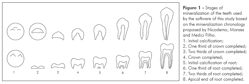
Panoramic radiographs were used for the evaluations. The radiographs of the patients were digitalized with a resolution of 254 dpi with an HP 4C scanner and Transparency Adapter (Hewllet Packard, Washington, USA). Assessment of dental age was performed with the aid of a software developed by the Discipline of Radiology, School of Dentistry of São José dos Campos, São Paulo State University (UNESP), using the table of mineralization chronology of permanent teeth among Brazilians proposed by Nicodemo, Moraes and Medici Filho (1974), which uses eight stages of dental mineralization (Figures 1 and 2). All teeth considered to be in the process of formation were used. According to those authors, the use of a greater amount of teeth increases the reliance of the calculation.
The panoramic radiographs were analyzed to observe the stages of mineralization of each tooth, and then the data were transferred into the software. After inputting the mineralization stage values for each tooth, the software gives the average dental age for each patient. Evaluation of the dental ages was performed by two examiners calibrated separately in two occasions with an interval of 15 days. The t test was used to check inter-examiner calibration.
Using the dental age values, graphs were drawn to compare chronological age (CA) and dental age (DA). Pearson's correlation test (p > 0.05) with descriptive analysis and diagrams of dispersion were carried out.
Comparison between the dental and chronological ages was made taking into account the percentage of patients with delayed or advanced dental age related to their chronological ages. Dental age could be delayed or advanced by 12 months, between 12 and 24 months, or above 24 months.
In 1974, Nicodemo, Moraes and Medici Filho established a dental mineralization chronology standard for Brazilians. The data obtained by these authors were used to produce the software used in the present study. Since there is a mineralization chronology standard for permanent teeth, there was no need for a control group.
Values with differences of up to 3 months were considered as statistically insignificant. Differences of up to 12 months were considered as representing slightly early or late ages, although they were regarded as being within the normal standards. Age differences over 12 months were considered to be outside normal standards. Age differences of more than 24 months were considered to be highly outside normal standards, meaning that children in that category are severely late or early regarding the average normal development standard.7
Results
Graphs 1, 2, 3 and 4 show the values of dental and chronological ages for the male and female subjects.
It can be seen in Graphs 1 and 2 that for both males and females the dental age values for most of the Down patients analyzed in the present research were considered to be within the normal standards (up to 12 months).
As to the differences between the dental and chronological ages, it can further be noted in Graph 1 that 24.53% (males) and 22.45% (females) displayed dental age delayed up to 12 months, and 3.78% of the males and 12.25% of the females displayed dental age delayed between 12 and 24 months.
It can also be noted in Graph 2 that 48.28% of the males and 38.77% of the females displayed dental age advanced up to 12 months, and 7.54% of the males and 16.32% of the females displayed dental age advanced between 12 and 24 months.
If the values pertaining to the up-to-12-month category (delayed or advanced) are added, 72.81% of the males and 61.22% of the females can be considered as having dental age within normal standard values. This means that approximately two thirds of the individuals studied here displayed normal age.
Graph 1 shows that 3.78% of the males and 4.09% of the females presented dental age delayed more than 24 months. Graph 2 shows that 15.09% of the males and 6.12% of the females presented dental age advanced more than 24 months. The added values of this category (more than 24 months) represent 18.87% of males and 10.21% of females with dental age significantly advanced or delayed, thus considered to be outside normal standards.
The dispersion diagrams represented in Graphs 3 and 4 show the sample distribution of dental age related to chronological age, with r = 0.92 for males and r = 0.89 for females. The regression line can be seen with 95% of confidence. The statistical analysis regarding inter-examiner calibration (t test) found p = 0.931 for males and p = 0.803 for females, meaning that there was no statistically significant inter-examiner difference.
Discussion
The results of the present study show a substantial variability in the dental age values as compared to the chronological age values, which were either higher or lower (delayed or advanced), and also differences between males and females. It is noteworthy that the estimated variations of the development parameters in relation to the dental and osseous ages as well as to puberal growth were deemed normal. The variation degree was evaluated in order to assess how different the chronological age was from the parameter evaluated.
Other authors' results were not presented because no reference to dental age in Down syndrome patients could be found in the literature. Hence, focus was placed on other parameters already studied related to dental development in order to make comparisons.
Regarding eruption, Mustacchi, Rozone8 (1990), Coelho, Loevy2 (1982) said that in Down patients the deciduous and permanent dentitions are sometimes late, and are more commonly variable than in the normal population. Mustacchi, Rozone8 (1990) mentioned that Down patients present a late dental eruption. Spitzer16 (1955) confirmed our results when he said that beyond the anomalies, dental malformation and eruption delays are severe in those affected by the syndrome. None of the reviewed studies cited an early dental development.
Santos14 (2004) also mentioned that the mineralization stages of the permanent teeth took place at different chronological times and that most stages of dental mineralization were slightly late, with differences of up to 12 months, which is in accordance with the results of our research. He also stated that, in some individuals, some stages were early. The author, therefore, suggests that a specific chronological table be elaborated for patients with Down syndrome.
In the present research, however, the observed percentages were unexpected. When we first suggested this study, we believed that, because Down patients present many kinds of dental development and formation alterations, dental age values different from normal ones were to be expected. This however did not happen as the dental age observed in this study for most of the Down syndrome patients was normal, with only small percentages of abnormality.
When dental age is compared to other development parameters, Sannomiya et al.13 (1998) mentioned that, in both male and female syndromic patients, there were no statistically significant differences between osseous and chronological ages.
This finding corroborated our results and led us to believe that these parameters have little influence in Down patients. According to Moraes7 (1998), it has been widely investigated and proven that the dental and osseous ages are closely related.
Conclusion
1. The dental age of two thirds of the male and female patients of the present study was predominantly advanced, and in one third of them, it was late.
2. The differences between the dental age and chronological age of two thirds of the male and female patients were only up to 12 months, which means that the majority of patients presented a normal development.
Acknowledgments
The authors of this research would like to thank Sandra Maria Herondina Coelho Ávila Aguiar and her supervisor, professors of the discipline of Radiology and of the Center for the Dental Treatment of the Mentally Disabled (CDTMD), School of Dentistry of Araçatuba, São Paulo State University. This research was supported by The State of São Paulo Research Foundation (FAPESP), through its Scientific Initiation scholarship n. 01/06866-2.
Received for publication on Aug 02, 2006
Sent for alterations on Jan 24, 2007
Accepted for publication on Apr 12, 2007
- 1. Coe DA, Matson JL, Russell DW, Slifer KJ, Capone GT, Baglio C et al Behavior problems of children with Down syndrome and life events. J Autism Dev Disord. 1999;29(2):149-56.
- 2. Coelho CRZ, Loevy HT. Odontological aspects of Down's syndrome. ARS Curandi Odontol. 1982;8(3):9-16.
- 3. Hayes A, Batshaw ML. Down syndrome. Pediatr Clin North Am. 1993;40:523-35.
- 4. Jensen GM, Cleall JF, Yip AS. Dentoalveolar morphology and developmental changes in Down's syndrome (trisomy 21). Am J Orthod. 1973;64(6):607-18.
- 5. Mariano MPK, Krahembull SMB, Magalhães MHC. Alterações sistêmicas de interesse odontológico na síndrome de Down. RPG Rev Pos Grad. 1999;6:218-21.
- 6. Moraes MEL, Bastos MS, Moraes LC, Rocha JC. Prevalência de cárie pelo índice CPO-D em portadores de síndrome de Down. PGRO-Pós-Grad Rev Odontol. 2002;5(2):64-73.
- 7. Moraes MEL, Medici Filho E, Moraes LC. Surto de crescimento puberal: relação entre mineralização dentária, idade cronológica, idade dentária e idade óssea - método radiográfico. Rev Odontol UNESP. 1998;27:111-29.
- 8. Mustacchi Z, Rozone G. Síndrome de Down: aspectos clínicos e odontológicos. São Paulo: CID; 1990.
- 9. Oliveira ACB, Ramos-Jorge ML, Paiva SM. Aspectos relevantes à abordagem odontológica da criança com Síndrome de Down. Rev do CROMG. 2000;15(2):24-9.
- 10. Pilcher ES. Dental care for the patient with Down syndrome. Downs Syndr Res Pract. 1998;5(3):111-6.
- 11. Rey SC, Fazzi R, Birman EG. Principais alterações craniofaciais em portadores de Síndrome de Down . Rev Fac Odontol FZL. 1991;3(1):59-64.
- 12. Ribeiro LMA, Jacob CMA, Pastorino AC, Kim CAE, Fomin ABF, Castro APBM. Evaluation of factors associated with recurrent and/or severe infections in patients with Down's syndrome. J Pediatr. 2003;79(2):141-8.
- 13. Sannomiya EK, Medici Filho E, Castilho JCM, Grasiozi MAOC. Avaliação da idade óssea em indivíduos portadores da sindrome de down, por meio de radiografias da mão e punho. Rev Odontol UNESP. 1998;27(2):527-36.
- 14. Santos LRA. Estudo radiográfico da cronologia da mineralização dos dentes canino, pré-molares e segundos molares em pacientes portadores de síndrome de Down [Dissertação de Mestrado]. São Paulo: Faculdade de Odontologia de São José dos Campos da UNESP; 2004.
- 15. Schwartzman JS. Síndrome de Down. 2Ş ed. São Paulo: Mackenzie; 2003.
- 16. Spitzer R. Radiological changes in teeth and skull in mental defectives. Br J Radiol. 1955;28(327):117-27.
- 17. Toscano MDP. Estágios cronológicos de desenvolvimento dentário em crianças portadoras de síndrome de Down. Estudo radiográfico [Dissertação de Mestrado]. Araraquara: Faculdade de Odontologia da UNESP; 1994.
Publication Dates
-
Publication in this collection
19 Oct 2009 -
Date of issue
Sept 2007
History
-
Reviewed
24 Jan 2007 -
Received
02 Aug 2006 -
Accepted
12 Apr 2007